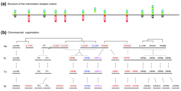
Figure 8.
Syntenic organization of classII cytokine receptor genes. (a) Diagram of the structures of the mammalian receptor chains, with the blue and green rectangles representing the S100A and S100B domains, the red rectangle the intracellular domain of the ligand binding chains, and the gray rectangle the intracellular domain of the non-ligand-binding chains and TF (after Renauld [57]). (b) Synteny between regions containing class II cytokine receptor genes in mammals and fish. Fat horizontal lines indicate chromosomes in the four species. The brackets above the human genes show evolutionary relationships between the paralogs. Vertical broken lines indicate suggested evolutionary relationships between the genes in the different species, based on the tree in Figure 9. Color coding of names: red = long intracellular domain; black = short intracellular domain; blue = no intracellular domain; and pink = intermediate length intracellular domain. Circled names indicate ligand binding chains. Round brackets denote groups of genes in cases where there are no clear orthologous relationships of individual members with genes in the other species.

