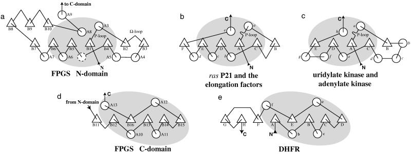
Figure 4.
Topology diagrams of (a) the N domain of FPGS, (b) p21-ras and G proteins, (c) ADK and uridylate kinase, (d) the C domain of FPGS, and (e) DHFR. The regions of structural similarity between the N domain of FPGS, the G proteins, and the kinases are indicated by the shaded areas in a–c. Likewise, structurally similar regions in the C domain of FPGS and DHFR are shaded in d and e. Secondary structure assignments for FPGS are: N domain; A1, 2–11; A2, 22–38; A3, 49–63; A4, 91–113; A5, 119–135; A6, 177–189; A7, 201–216; A8, 260–280; A9, 284–294; B1, 40–45; B2, 67–72; B3, 83–86; B4, 139–144; B5, 160–164; B6, 194–198; B7, 218–222; B8, 227–234; B9, 237–244; B10, 247–254; C domain; A10, 320–332; A11, 351–359; A12, 390–402; A13, 412–425; B11, 302–305; B12, 309–313; B13, 336–341; B14, 361–365; B15, 386–388; B16, 406–410.

