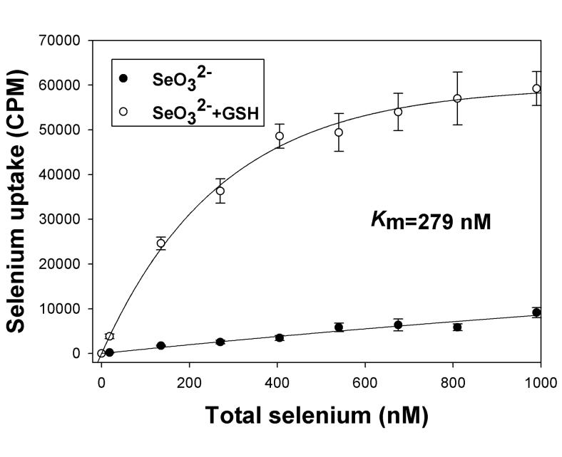
Figure 1. Glutathione reduction stimulates selenium uptake in HaCat cell model.
Uptake of selenite (SeO32−) and reduced selenium (SeO32− + GSH) was assessed after incubation of radiolabeled selenium with cells in DPBS for 60 minutes. The concentration of selenium was varied without changing the specific activity of the radioisotope as described in Methods. A representative experiment is shown, taken from two independent reproducible experiments (n=6). The data is shown as means with standard deviation plotted as error of three independent culture replicates.

