Figure 5.
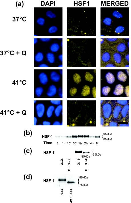
The effect of HS on HSF-1 activation in Caco-2 monolayers. The effect of HS on HSF-1 cytoplasmic-to-nuclear translocation as determined by immunofluorescent antibody labeling as described in Materials and Methods. a: After appropriate treatment, Caco-2 monolayers were fixed and co-stained for HSF1 by using specific antibodies and corresponding secondary antibodies conjugated with fluorescent probes. Nuclei were visualized with DAPI (blue). Heat exposure: 41°C for 1 hour; quercetin (100 μmol/L). b: Time-course effect of HS-induced HSF-1 activation in Caco-2 monolayers. Caco-2 monolayers were treated with HS (41°C) for increasing time points (0 to 8 hours) and changes in the HSF-1 expression in the nuclear fraction were assayed by Western blot analysis. c: Effect of HSF-1 inhibitor quercetin on heat-induced increase in HSF-1 activation. Caco-2 monolayers were treated with 41°C alone or with quercetin (100 μmol/L) for the 1-hour experimental period. Subsequently, HSF-1 protein levels in nuclear fraction were determined by Western blot analysis as described in the Material and Methods. Quercetin treatment at 41°C resulted in an inhibition of HS-induced increase in HSF-1 protein expression. d: Effect of dephosphorylating enzyme alkaline phosphatase on heat-induced increase in HSF-1 phosphorylation. Caco-2 protein lysate (after a 1-hour exposure to 41°C) was treated with alkaline phosphatase. Alkaline phosphatase treatment resulted in disappearance of higher molecular weight forms of HSF-1. The experiment was repeated three to four times. Scale bar = 20 μm.

