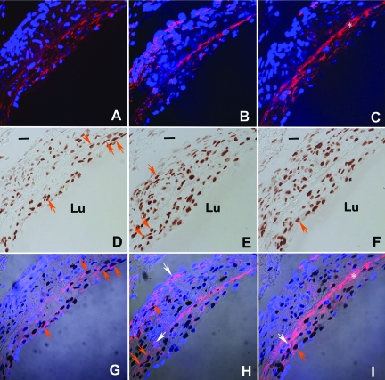
Figure 1.
Analysis of 2 week neointima lesion:proliferating cell type. A–C: Confocal image of smooth muscle myosin heavy chain (A, red) and CD45-positive cells (B, red) and DAPI nuclear staining (blue). C: vWF (red) and DAPI nuclear staining (blue). Because of injury to the endothelium, platelets (which are vWF-positive) accumulate along the lining of the vessel. This is reflected as a bright, red, anuclear, fluorescent band indicated by the *. Given that platelets do not have a nucleus, their positive fluorescence is not an indication of an endothelial cell. D–F: Bright-field image of PCNA-positive cells (brown). G–I: Merged image: orange arrows indicate example of PCNA-positive smooth muscle cells (G), leukocytes (H), or endothelial cells (I). The identical cells in D, E, and F are indicated by orange arrows. Example of nonproliferating leukocytes (H) and endothelial cells (I) are indicated by white arrows. Note: in the adventitia there is a predominance of CD45-positive cells (H) with a paucity of smooth muscle cells (G) and endothelial cells (I). Bar = 20 μm, Lu, lumen.

