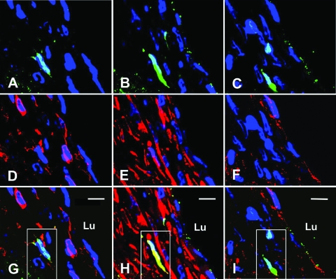
Figure 2.
Identification of bone marrow-derived cells within the neointima 2 weeks after grafting. A–C: Three adjacent sections imaged by confocal microscopy after immunofluorescent detection of GFP-positive cells (green) and stained with DAPI nuclear stain (blue). D–F: Red fluorescence indicates CD45 (D), smooth muscle myosin heavy chain (E), or vWF (F) of same sections pictured in A to C, respectively. G–I: Merged image of GFP fluorescence (A–C) and red fluorescence images (D–F). Note the GFP-positive cell expressing smooth muscle myosin heavy chain appears yellow (outlined by a white box) in H. This same cell, since it does not express CD45 or vWF, appears green in G and I, respectively. Bar = 10 μm, Lu, lumen.

