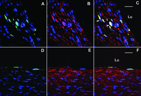
Figure 3.
Identification of bone marrow-derived cell at 8 weeks (A–C) and 16 weeks (D–F) after grafting. A,D: Confocal image of GFP-positive cells (green) and DAPI nuclear stain (blue). B,E: smooth muscle myosin heavy chain-positive cells (red) and DAPI nuclear stain (blue). C: Merged image of 8-week lesion demonstrating that GFP-positive cells also express smooth muscle myosin heavy chain (C, white arrow). F: Merged image of 16-week lesion demonstrating GFP-positive cells outline the lumen (and stain positive for vWF (data not shown)), but no smooth muscle myosin heavy chain expressing cells expresses GFP. Bar = 20 μm, Lu, lumen.

