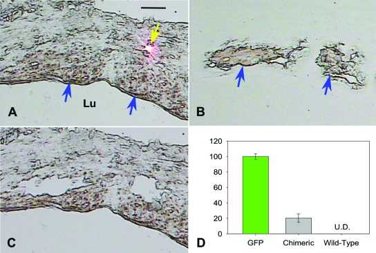
Figure 5.
DNA determination of origin of smooth muscle cells at 16 weeks after grafting. A: Bright-field image of section before capture. Smooth muscle cells were identified with immunohistochemistry stain (brown, blue arrow) and captured by laser (pink dot indicates laser (yellow arrow)). B: Captured smooth muscle cells on a transparent cap. C: Void within neointima lesion left by captured smooth muscle cells. Bar = 20 μm, Lu, lumen. D: Real-time PCR determination of percentage of GFP-positive cells, as determined by copy number of GFP vs. β-actin, among captured smooth muscle cells isolated from chimeric mice. Shown as controls are results of real-time PCR of smooth muscle cells isolated from a GFP and wild-type mice (U.D., undetectable).

