
Figure 1.
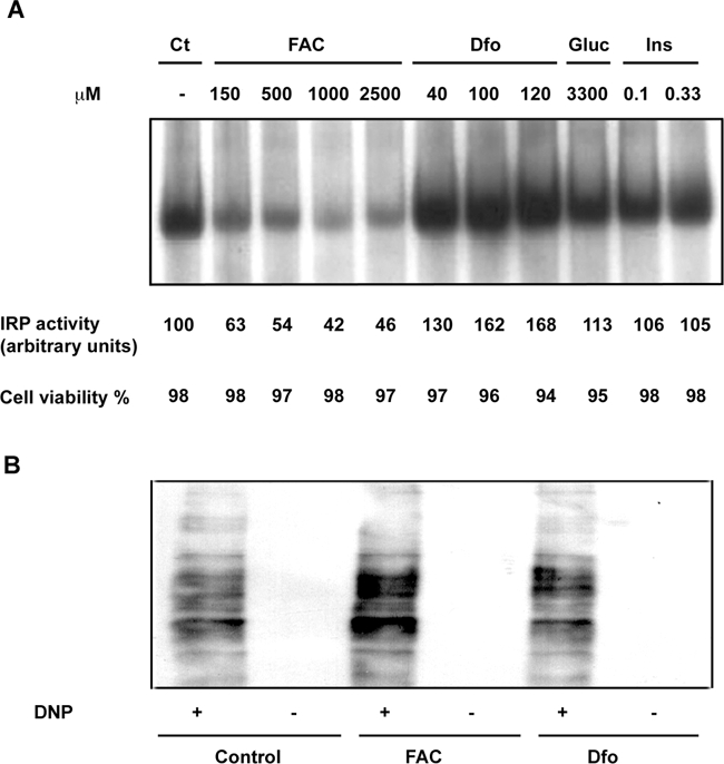
Effect of iron manipulation on IRP activity, cell viability, and oxidative stress. A: IRP activity assay. Cytoplasmic extract of HepG2 exposed to increasing concentrations of FAC, Dfo, glucose (33 mmol/L, Glu), and insulin (0.33 μmol/L, Ins), were incubated with an excess of a 32P-labeled iron-responsive element probe; RNA-protein complexes were separated on nondenaturing 6% polyacrylamide gels and revealed by autoradiography. The corresponding percent cell viability, evaluated by Trypan blue, is indicated at the bottom. B: Protein carbonylation by Oxyblot in cells treated with FAC, Dfo, and untreated. DNP+, derivatization to 2,4-dinitrophenylhydrazone; DNP−, negative control.