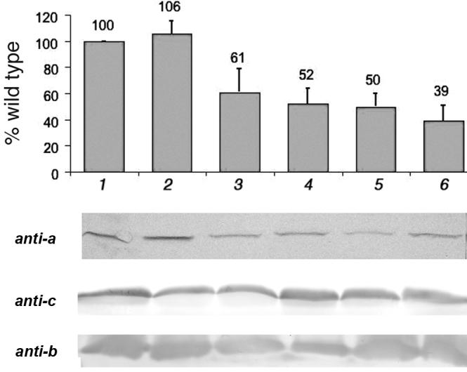
Fig. 2.

Quantification of subunit a in membrane vesicles by immunoblotting. Membrane vesicles were solubilized by dodecyl sulfate, separated by SDS-PAGE, blotted, and probed with polyclonal anti-a antibodies. The bands were measured by densitometric analysis, and the means of 3-5 experiments are shown, with standard deviations. The y-axis is the percent of the wild type value. All samples were from DK8/pFV2 with the indicated mutations: (1) wild type. (2) P204T/R210Q/Q252R. (3) P204T/R210Q/Q252K. (4) R210Q/Q252R. (5) R210Q/Q252K. (6) R210Q. Below, representative blots are shown using anti-a, anti-c and anti-b antibodies.
