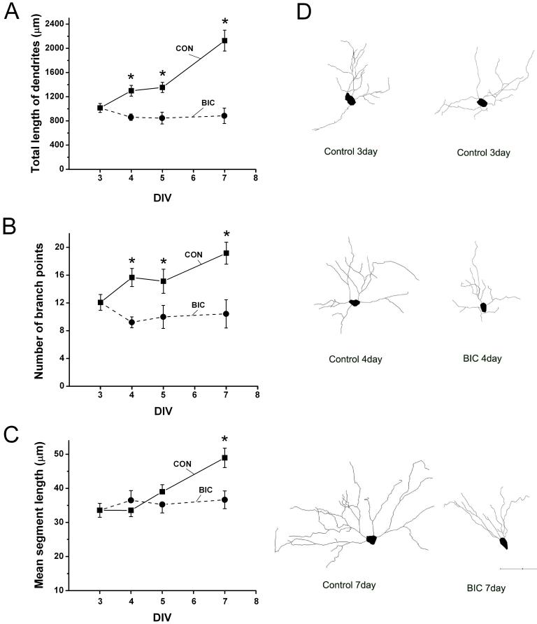
Fig. 4.
Chronic disinhibition suppresses the growth of CA1 hippocampal pyramidal cell dendrites. (A-C) Between 3 and 7 days in vitro (DIV), the dendrites of CA1 pyramidal cells in control slices gradually increased in total length, number of branch points and mean segment length. However in bicuculline treated slices, dendrites did not increase in total length (A) or average length of individual segments (C). The number of dendritic branch points appears to decrease during bicuculline treatment (B) but this effect was not statistically significant. (D) Representative Neurolucida reconstructions of dendrites of YFP positive CA1 pyramidal neurons at selected times in vitro. The soma and basilar dendrites of these cells are shown and clearly illustrate that neurons treated with bicuculline have simpler dendritic arbors compared with controls at the same time. Number of neurons reconstructed: Control n= 6-13, Bicuculline n= 7-14. Scale bar = 100 μm. Pairwise comparison of control versus bicuculline treated dendrites on DIV 4, 5 and 7 were significantly different as indicated by * (p ≤ 0.05).

