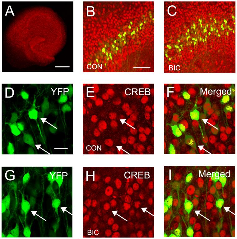
Fig. 7.
Expression of the transcription factor CREB is unaltered by chronic disinhibition. (A) Confocal image illustrating the typical pattern of CREB expression in a hippocampal slice culture. (B and C) Very similar CREB expression patterns are observed in hippocampal area CA1 in control and bicuculline treated slice cultures. YFP positive pyramidal cells are also shown in these images. (D-I) The nuclear localization of CREB in YFP positive CA1 pyramidal cells is illustrated in merged image. (G-H) The pattern and degree of CREB expression appears unaltered after 4 days of bicuculline treatment. The majority of CREB positive - YFP negative cells are CA1 pyramidal cells that do not express YFP. Arrow heads indicate co-localization of CREB and YFP signal. Calibration bars: A = 500 μm. B and C = 100 μm. D - I = 20 μm.

