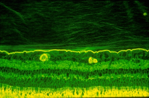
Fig. 2.
Vitreo–retinal interface. Imunohistochemical studies of the monkey vitreo–retinal interface employed fixation in 4% paraformaldehyde and staining with fluorescein-conjugated ABA lectin. The retina is at the bottom and the vitreous is at the top of this image. The intensely-stained, horizontal linear structure is the internal limiting lamina (ILL) of the retina. Above the ILL is the posterior vitreous cortex. The lamellar structure is clearly evident. Effective magnification ∼400x. (Courtesy of Greg Hageman, Ph.D.)

