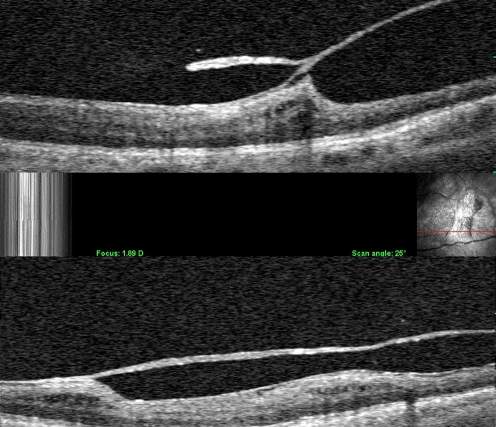
Fig. 4.
Clinical vitreoschisis. Combined OCT/SLO imaging detected a split in the posterior vitreous cortex. In these two cases, the outer layer of the split posterior vitreous cortex remains adherent to the retina. The point where the two layers re-join into one full-thickness layer is often the site of significant traction upon the retina. Studies have shown that about half of patients with macular hole and macular pucker have vitreoschisis

