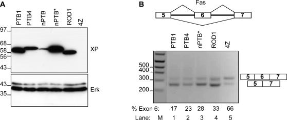
Figure 6. Splicing repressor activity of nPTB and ROD1.
A splicing reporter comprising FAS exons 5-6-7 [31] was cotransfected with expression constructs for PTB1, PTB4, nPTB* and ROD1 into L cells. PGEM4Z was used in the negative control. FAS exon 6 contains a single PTB binding site that mediates exon skipping [31]. A) Western blot probed with anti-Xpress (top) and anti-ERK (lower panel) antibodies. Size markers (kDa) are indicated to the left. Comparable levels of expression were obtained for PTB, ROD1 and nPTB from the codon-optimized nPTB* construct, but not from the original nPTB construct. B) RT-PCR analysis of FAS construct splicing. Products were separated by agarose gel electrophoresis. Lane M, size markers (bp), indicated to the left. Lanes 1-5, PTB1, PTB4, nPTB*, ROD1 and pGEM4Z respectively. Numbers immediately below each lane indicate the percentage of spliced product that includes exon 6. Both nPTB* and ROD1 were able to induce similar levels of exon skipping as PTB. Note that nPTB was not included in this panel because the protein is not adequately expressed (panel A).

