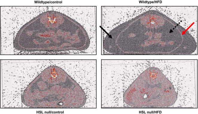
Figure 2. Subcutaneous and visceral adipose tissue stores are decreased in HSL null mice.
Representative CT scan images displaying body fat distribution in 10–11 months old female wildtype and HSL null mice fed either a control diet or a HFD for 6 months (n = 7). The filled black arrow and the dotted black arrow indicate the location of the subcutaneous and visceral WAT, respectively. The red arrow indicates the location of the muscle layer separating the two adipose tissue depots.

