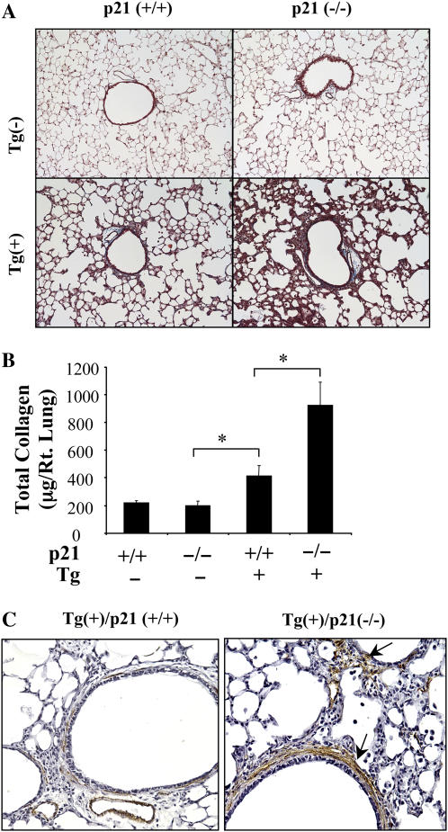
Figure 3.
Role of p21 in the TGF-β1–induced fibrosis. The collagen content of lungs from Tg (−) and Tg (+) mice with (+/+) and (−/−) p21 loci were compared using Mallory's trichrome (A; original magnification: ×10) and Sirchol collagen evaluations (B) after 2 weeks of dox induction. (C) Immunohistochemical localization of α-smooth muscle actin–positive cells (arrows) after 2 weeks of dox induction (original magnification: ×15). A and C are representative of a minimum of three similar evaluations. In B, each value represents the mean ± SEM of evaluations in a minimum of six mice (*P < 0.05).

