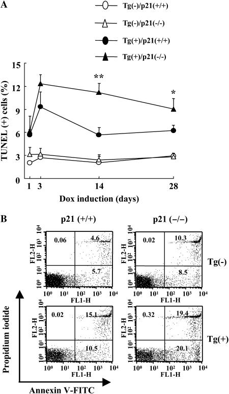
Figure 5.
p21 regulation of TGF-β1–induced apoptosis. After dox induction, lungs were obtained from Tg (−) and Tg (+) mice with (+/+) and (−/−) p21 loci. DNA injury and cell death were evaluated with TUNEL stains and time kinetic changes for the indicated dox induction period were illustrated (A). After 2 weeks of dox induction, cells were isolated from both lungs and fluorescence-activated cell sorter analysis was undertaken using annexin V and propidium iodide staining (B). In B, the values represent the mean ± SEM of evaluations in a minimum of six mice (*P < 0.05, **P < 0.01).

