
Figure 3. Primary human microglia and macrophages, but not astrocytes show increased [3H]DAA1106 binding on activation.
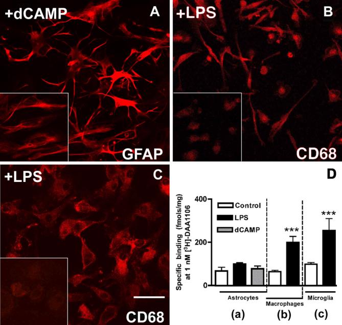
A, Primary human embryonic astrocytes activated with dB-cAMP show changes in morphology with the appearance of spindle shaped processes and increased GFAP staining compared to non-activated cultures (inset).
B, Primary human macrophages activated with LPS show changes in morphology from a rounded shape (inset) to spindle shaped with increased CD68 staining in non-stimulated cultures.
C, Primary human embryonic microglia were activated with LPS for 48 hrs show increased CD68 staining compared to non-activated cultures (inset).
D, [3H]DAA1106 specific binding (fmols/mg mitochondrial protein) was significantly higher in mitochondrial preparations obtained from both macrophages (b) and microglia (c) activated with LPS (black bars) compared to unactivated controls (white bars) and astrocytes (a) with or without activation. Data was analyzed using ANOVA, n=3 in each group, ***p<0.0001.