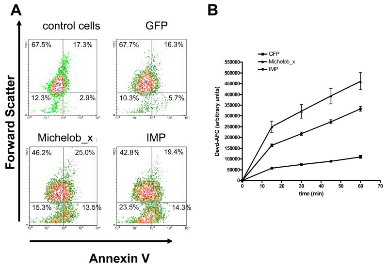
Figure 4.
Expression of IMP or Michelob_x causes apoptosis in C6/36 cells. A, AnnexinV staining (X-axis) versus forward scatter (Y-axis) of untransfected (control) cells or cells transfected with constructs expressing GFP, Michelob_x, or IMP, as analyzed by flow cytometry. Percentages are given for each quadrant. Each graph represents analysis of 10,000 cells. B, caspase activity in lysates from cells expressing GFP, Michelob_x, or IMP as determined by liberated AFC fluorescence over 60 min incubation with the caspase substrate Ac-DEVD-AFC. The data shown represent the combined results from three independent transfections.

