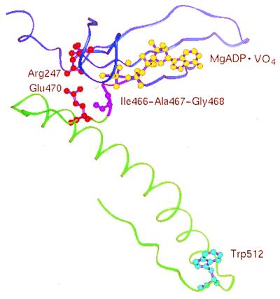
Figure 8.
The neighborhood of the Glu–Arg salt-bridge, and the “sensitive” tryptophan. The entire structure of the truncated myosin head from Dictyostelium ligated at the active site with MgADP⋅VO4 was solved by Smith and Rayment (4). To get an idea of how this neighborhood might look in the smooth muscle heavy meromyosin used in this work, the residues located by Smith and Rayment have been labeled as though they were the conserved homologs of the smooth muscle sequence (17). Backbone atoms of the 235–278 and 458–465 sequences of the heavy chain are colored blue, and the 469–521 sequence is colored green. Residues Ile-466–Ala-467–Gly-468 are colored magenta. Residues 247 (in red), 470 (in red), and 512 (in cyan) are shown as balls and sticks, as is the analog MgADP⋅VO4 (in yellow), simulating MgADP⋅Pi.

