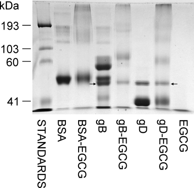
FIG. 7.
Envelope glycoproteins gB and gD form macromolecular complexes in the presence of EGCG. EGCG (545 μM) was incubated with recombinant purified gB or gD or with purified BSA for 24 h and then analyzed by SDS-PAGE, followed by Coomassie blue staining. All of the bands in lanes with gB and gD (with or without EGCG) were analyzed by tandem mass spectrometry (Mass Spectrometry Facility, New York State Institute for Basic Research). Arrows indicate the presence of endochitinase either alone in the gD lanes or mixed with gB.

