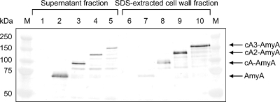
FIG. 2.
Western blot analysis of AmyA and cA-AmyA fusion proteins from the supernatant fraction (lanes 1 to 5) and the SDS-extracted cell wall fraction (lanes 6 to 10) of genetically modified L. lactis IL 1403 cells. Lanes: M, marker proteins with molecular masses indicated; 1 and 6, cells harboring pCUS; 2 and 7, cells harboring pMCS-AmyA; 3 and 8, cells harboring pCA-AmyA; 4 and 9, cells harboring pCA2-AmyA; 5 and 10, cells harboring pCA3-AmyA. The samples were separated by SDS-polyacrylamide gel electrophoresis with an 8% gel and stained with rabbit anti-AmyA serum, followed by secondary goat anti-rabbit IgG conjugated with alkaline phosphatase.

