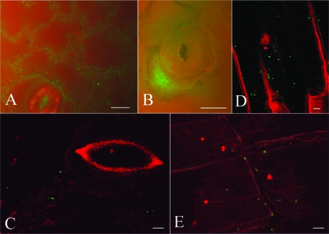
FIG. 3.
Visualization of fluorescent cells and natural transformation of the reporter strain A. baylyi BD413(rbcL-ΔPaadA::gfp) in situ on decaying tobacco tissues. To demonstrate fluorescence, A. baylyi strain BD413(rbcL-aadA::gfp), constitutively expressing GFP, was inoculated onto defrosted leaves and incubated for 5 days. GFP fluorescence showed single cells and microcolonies of A. baylyi in the interstices of epidermal cells (A) and between a stoma and the border of epidermal cells (B). (C to E) In situ detection by CLSM of natural transformation of the reporter strain BD413(rbcL-ΔPaadA::gfp) exposed to externally added DNA on decaying tissues of tobacco. In the experiment whose results are shown in panel C, leaf tissue was supplemented with purified pCLT DNA, while in the experiments whose results are shown in panels D and E, root tissue was supplemented with cell lysates of E. coli XL1-Blue(pCLT). The images show bacterial transformants on a leaf surface near a stoma (C) or scattered along the root tissues (D and E). Bars represent 10 μm.

