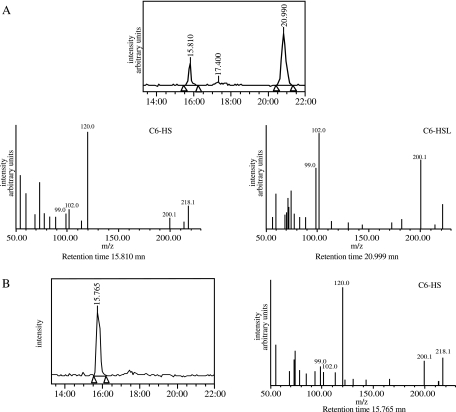
FIG. 3.
QsdA lactonase activity. E. coli strains DH5α(pME6032) and DH5α(pSU40) were incubated in pH 6.5 buffered LBm medium with 50 μM C6-HSL for 24 h. The medium was analyzed at 0 and 24 h by HPLC-mass spectrometry. Under the experimental conditions used, C6-HS (molecular weight, 217) and C6-HSL (molecular weight, 199) had retention times of 15.8 and 21 min, respectively, and mass spectra were composed of the following main fragments: m/z = 218, 200, and 120 for C6-HS, and m/z = 200, 102, and 99 for C6-HSL. (A) Spontaneous degradation of C6-HSL in aqueous medium. (B) DH5α(pSU40) after 24 h of incubation. A single peak at a retention time of 15.8 min is visible on the HPLC spectrum, which is identified as C6-HS. C6-HSL has completely disappeared from the medium. The formation of C6-HS, correlated with the absence of HSL in the medium, is indicative of a lactonase activity.

