FIG. 6.
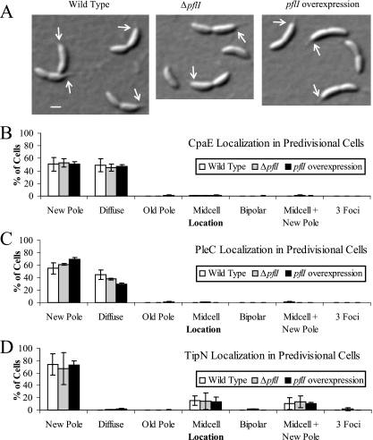
Localization of polar markers CpaE, PleC, and TipN is not significantly affected by altered PflI abundance. (A) Polar morphogenesis does not appear to be affected by pflI. Differential interference contrast images of wild-type (CJW1388; left panel), ΔpflI (CJW2188; center panel), and pflI-overexpressing (CJW2189; right panel) cells show normal cellular asymmetry and stalk formation. Arrows point to the base of the stalk. Scale bar, 1 μm. (B) Localization of CpaE-yellow fluorescent protein was determined in synchronized predivisional cells of strains PV418 (wild type), CJW2188 (ΔpflI), and CJW2189 (with pflI overexpression in xylose-containing medium) by fluorescence microscopy. The localization of a fluorescent focus, if any, was assigned to the category of diffuse, new pole, old pole, midcell, bipolar, midcell plus new pole, or three foci. Cells were considered to have diffuse localization if no focus was present. Cells were determined to have a focus at the new pole if the focus was present at the pole opposite the stalk or at the pole closer to the division site (when no stalk was visible) because C. crescentus cells divide off-center, slightly closer to the new pole. Cells with old-pole localization had a focus at the stalked pole or at the pole farther from the division site. Cells with midcell localization had a focus at the division site. Cells with foci at both poles were called bipolar. Cells with two foci (one at the division plane and the other at the new pole, as described above) were assigned to the midcell-plus-new-pole category. Cells with foci at both poles plus the division plane were identified as having three foci. (C) Localization of PleC-monomeric yellow fluorescent protein was visualized by fluorescence microscopy using synchronized predivisional-cell populations of CJW686 (wild type), CJW2184 (ΔpflI), and CJW2185 grown in xylose (with pflI overexpression). Fluorescent foci were categorized as described for panel B. (D) Predivisional cells of CJW1406 (wild type), CJW2186 (ΔpflI), and CJW2187 grown in xylose (with pflI overexpression) were observed by fluorescence microscopy to determine TipN-GFP localization. Fluorescent foci were categorized as described for panel B.

