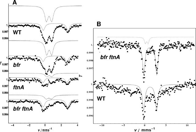
FIG. 4.
Mössbauer spectra of Erwinia chrysanthemi 3937 cells measured at 77 K (A) and 2 K (B). T represents the relative transmission, and v represents the energy scale measured as the velocity in mm/s. Genotypes of the different strains are indicated on the spectra. Cells were grown to an OD600 of 0.8 and incubated for 120 min with 5 μM 57Fe-100 μM DHBA. (A) Each spectrum is characterized by two quadrupole doublets, the parameters of which are listed in Table 4. The dashed gray line corresponds to the least-square fits of ferric high-spin iron to the experimental spectra, and full gray lines correspond to ferrous high spin. (B) Mössbauer spectra from cells of the bfr ftnA double mutant and the wild-type (WT) strain of E. chrysanthemi 3937 measured at 2 K. In both spectra, a ferrous high-spin component is observed (full gray lines). In the double mutant, the ferric ion doublet is still visible at 2 K (dashed gray line), whereas in the wild-type strain, this component broadens at temperatures below 4.3 K due to magnetic splitting, and the doublet disappears. The magnetic splitting is not resolved due to relaxation effects and was not fitted.

