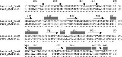
FIG. 2.
Pairwise alignment of the sequences of LuxG from P. leiognathi TH1 isolated in the present study (isolated_luxG) and LuxG previously reported (accession number AAA25621). The conserved residues are indicated by asterisk marks (*). Boldface letters represent the residues identical to those of E. coli Fre. Based on the structure of E. coli Fre (15), the flavin reductase can be divided into two domains; the N-terminal domain that binds flavin (indicated by “F”) and the C-terminal domain that probably binds NAD(P)H (indicated by “N”). Based on the structure of Fre (15), the secondary structure elements are shown above the text (β strands are indicated by arrows and α helices are indicated by solid blocks).

