FIG. 7.
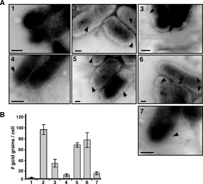
Immunoelectron microscopy detects Tzs on the A. tumefaciens cell surface. Strains C58, CB1002 (ΔvirB2), CB1005 (ΔvirB5), and CB1008 (ΔvirB8) and complemented variants were cultivated on AB minimal medium in the absence or in the presence of AS for virulence gene induction, followed by immunoelectron microscopy with Tzs-specific primary antibody and 10 nm gold-labeled secondary antibody. (A) Representative images of transmission electron micrographs; arrowheads point to gold grains on the cell surfaces of samples as follows: 1, C58 without AS; 2, C58 with AS; 3, CB1002; 4, CB1005; 5, CB1002 pTrcB2; 6, CB1005 pTrcB5; 7, CB1008. The contrast was increased to visualize the outline of cells for the purpose of presentation, but counting was conducted with reduced contrast settings that allowed the visualization of grains in even more heavily stained regions of the cells. Bars, 100 nm. (B) Quantification of results of the transmission electron microscopy analysis of Tzs on the cell surface; numbering of bars as for panel A. We counted 10 cells each from three independent induction experiments for each strain (total of 30 cells), and error bars show the standard deviations.

