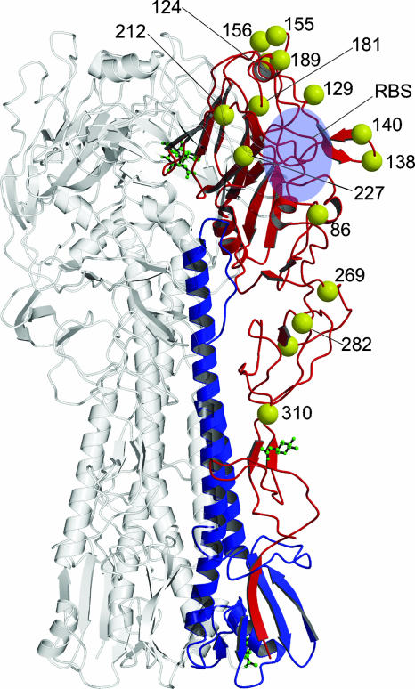
FIG. 2.
Ribbon diagram of the trimeric HA molecule with the mutations shown as yellow spheres. The majority of the polymorphic sites (Table 2) are exterior residues, with the exception of residues 94, 174, and 263 (not shown). The RBS is indicated with a blue oval. The reference monomer is drawn in color, and the other two monomers are shown in gray. In the reference monomer, polypeptide HA1 is in red, polypeptide HA2 is in blue, and the polysaccharides are drawn in ball-and-stick form in green. The diagram was prepared with the program MOLSCRIPT (23) and Raster3D (30).

