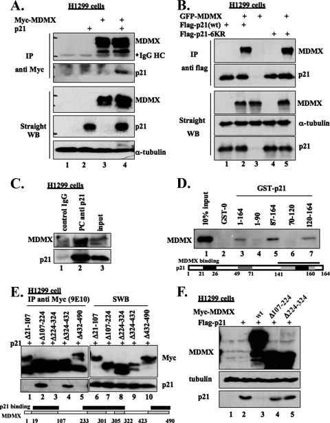
FIG. 3.
p21 and MDMX interact in cells and in vitro. (A) p21 interacts with MDMX in cells. H1299 cells were transfected with the p21 plasmid alone or together with c-myc-MDMX plasmid. The transfected cells were treated with 10 μM MG132 for 16 h and harvested at 48 h posttransfection and lysed. The cell lysates were immunoprecipitated using monoclonal anti-myc (9E10) antibody (300 μg/for each sample) or directly loaded for straight WB (50 μg/sample). The detected proteins are indicated on the right. HC, heavy chain. (B) Reciprocally, MDMX interacts with both wild-type and lysine mutant p21. The Flag-tagged wild-type p21 (wt) or p21-6KR (a p21 mutant in which all lysine residues of p21 were replaced by arginines) was transfected alone or together with GFP-MDMX, in H1299 cells, and treated as in panel A. The cell lysates were immunoprecipitated with monoclonal anti-Flag antibody followed by WB analyses, as indicated on the right. (C) Endogenous p21 complexes with MDMX. H1299 cell lysate (400 μg) was immunoprecipitated with polyclonal anti-p21 antibody or control immunoglobulin G (IgG) and probed for p21 and MDMX. An aliquot of the same cell lysate (80 μg) was used for input. (D) MDMX physically associates with the C terminus of p21 in vitro. Preparation of GST-fusion protein beads and purification of His-MDMX were described previously (20). His-MDMX (100 ng) was incubated with beads conjugating 500 ng GST-0 (GST only) or GST-fused full-length p21 or fragments of p21 in lysis buffer. Thirty minutes after incubation at room temperature, the mixtures were washed with lysis buffer once, SNNTE buffer twice, and lysis buffer again. The samples, together with 10% His-MDMX input, were resolved by SDS-PAGE, followed by straight WB (SWB) against MDMX (8C6). (E) Mapping of p21-binding domains on MDMX in cells. H1299 cells were transfected with Flag-p21 alone or together with myc-tagged wild-type MDMX or MDMX deletion mutants. The cells were treated and lysed as in panel A. The lysates were immunoprecipitated with monoclonal anti-myc antibody (9E10), followed by WB analyses, as indicated on the right. (F) The MDMX deletion mutants that do not bind to p21 lack the ability to decrease p21 levels in cells. H1299 cells were transfected with Flag-p21 alone or together with myc-tagged wild-type MDMX or MDMX deletion mutants. The transfected cells were harvested at 48 h posttransfection and lysed. The cell lysates (50 μg/sample) were used for straight WB, as indicated on the left.

