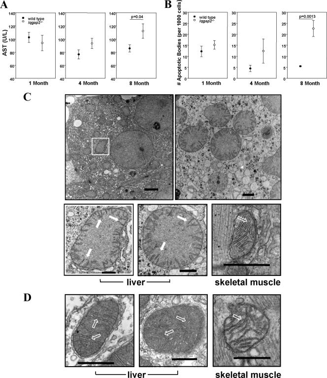
FIG. 2.
Age-dependent hepatopathy and mitochondrial damage in Iqgap2−/− mice. (A) Sera from mice in three age groups (1, 4, and 8 months old; n = 10 mice per age group) were analyzed for the quantification of AST. (B) Liver sections (n = 3 per age group) were analyzed for apoptotic cells by TUNEL staining. Results shown in panels A and B are means ± SEM; statistically significant P values are shown. (C and D) Transmission electron micrographs from livers and skeletal muscles of 12-month-old Iqgap2−/− mice (low and high magnifications) (C) or wild-type mice (high magnification) (D) demonstrate an edematous mitochondrial matrix with collapsed cristae (solid arrows) restricted to Iqgap2−/− mitochondria; hollow arrows indicate normal cristae. Note the normal architecture of mitochondria from skeletal muscles of both genotypes. Structural defects are representative of those seen in 8- and 12-month-old mice (n = 3 per group), with comparable but less-extensive defects seen in 4-month-old Iqgap2−/− mice. Size bars represent 2 μm (magnification, ×4,800 [C, top left panel]), 500 nm (magnification, ×18,500 [C, top right panel]), and 200 nm (magnification, ×49,000 [C, bottom panels, and D]). The white box denotes an Iqgap2−/− mitochondrion shown magnified at ×49,000 in the image below.

