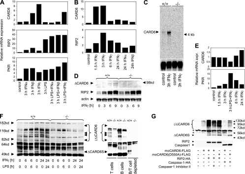
FIG. 3.
Regulation of CARD6 expression in BMDMs and splenocytes by IFNs and LPS. (A) Relative CARD6, RIP2, and PKR mRNA expression in BMDMs. BMDMs were stimulated for 3 h with 100 U ml−1 IFN-α or IFN-β, 50 ng ml−1 IFN-γ, or 100 ng ml−1 LPS or the indicated combinations of IFNs and LPS. RNA expression was measured by quantitative real-time PCR analysis. Results shown are mRNA levels relative to mRNA levels in unstimulated controls. (B) CARD6 mRNA induction by IFN-γ is rapid and transient. BMDMs were stimulated for the indicated times with 100 U ml−1 IFN-γ, IFN-α, or IFN-β. Relative mRNA expression was determined as for panel A. (C) Confirmation of CARD6 mRNA induction. CARD6+/+ and CARD6−/− BMDMs were left untreated or stimulated with IFN-γ for 3 h, and CARD6 RNA expression was detected by Northern blotting. (D) Upregulation of the ΔCARD6 protein variant in WT BMDMs. CARD6+/+ and CARD6−/− BMDMs were stimulated with IFN-γ for the indicated times, and extracts were subjected to Western blotting to detect CARD6, RIP2, and actin. (E) Relative CARD6 and PKR mRNA expression in splenocytes. Freshly isolated CARD6+/+ splenocytes were stimulated with IFN-γ in the absence or presence of LPS for the indicated times, and relative mRNA expression was determined as for panel A. (F) CARD6 protein expression in splenocytes. Left panel, CARD6+/+ splenocytes were incubated with IFN-γ in the absence or presence of LPS for the indicated times, and the expression of the (Δ)CARD6 and ΔCARD6S proteins was determined by Western blotting. Right panel, base levels of CARD6 protein expression were determined in unstimulated WT splenic T cells and B cells and in WT splenocytes depleted of T and B cells. Protein extracts from CARD6−/− splenocytes are included as controls in both panels. (G) Proteolytic CARD6 degradation is not altered by a mutation in a putative caspase-1 cleavage site or by caspase-1 inhibition. 293T cells were transfected with empty vector or the indicated CARD6 expression constructs in the absence or presence of expression constructs for HA-tagged RIP2 or caspase-1. Where indicated, cells were also treated with caspase-1 inhibitor II (Calbiochem) at a final concentration of 10 μM. Cells were lysed at 36 h posttransfection, and expression levels of exogenous CARD6 (upper panel) or HA-tagged RIP2 and caspase-1 (lower panel) were analyzed using anti-moCARD6 antibody or anti-HA antibody, respectively. Results shown are from one trial, representative of at least three (A, B, D, and E) or two (F and G) independent experiments. *, cross-reactive bands.

