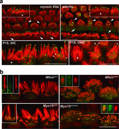
FIG. 5.
Both myosin XVa and whirlin localize to the tips of myosin VIIa-deficient stereocilia, but the whirlin labeling pattern persists abnormally during bundle development. (a) Myosin XVa and whirlin localized (green) to the tips of stereocilia of myosin VIIa-deficient inner (arrows), outer (arrowheads), and complemented hair cells at P7. Insets show representative inner hair cells at a higher magnification; complemented hair cells are indicated by asterisks. Interestingly, whirlin could still be observed in the tips of all myosin VIIa-deficient stereocilia at P15 (bottom, inner hair cells [IHC] and outer hair cells [OHC]) while being restricted to the most lateral stereocilia of complemented inner hair cells (*). Note the elevated levels of whirlin immunoreactivity in the cuticular plates of cells lacking myosin VIIa (pixel intensity values in arbitrary units for mutant outer hair cells are 27.24 ± 2.88, [n = 34] for complemented outer hair cells and 16.70 ± 2.09 [n = 27] for mutant outer hair cells [P = 2.5 ×10⁻²² by t test]). (b) In mice heterozygous for whirler (Whrnwi/+) and shaker2 (Myo15sh2/+) alleles, myosin VIIa (green) is present along the length of the most lateral (tallest) stereocilia (top, left inset) and at the tips of the shorter stereocilia (bottom, left inset). In the abnormally short stereocilia of mice lacking whirlin (Whrnwi/wi) and myosin XVa (Myo15sh2/sh2), myosin VIIa localizes at the tips. Insets show isolated stereocilia at a high magnification; merged images on the left show a green channel in the middle and a red channel on the right. Actin filaments in stereocilia were counterstained with rhodamine/phalloidin (red). Scale bars, 10 μm.

