FIG. 5.
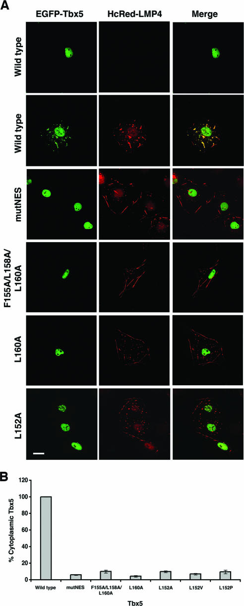
Subcellular localization of Tbx5 NES mutants in the presence of LMP4. (A) COS-7 cells were cotransfected without or with HcRed-LMP4 and the indicated EGFP-Tbx5 expression vectors. After transient expression, cells were fixed and imaged using a Zeiss 510 Meta scanning confocal microscope with a Plan Apochromat 63× objective/1.4 numerical aperture oil differential interference contrast lens. The left column shows the localization of the EGFP-Tbx5 proteins, the middle column shows the localization of the HcRed-LMP4 protein, and the right column shows both proteins together in a merged image. When expressed alone, wild-type Tbx5 is detected only in the nuclei, while when coexpressed with LMP4, wild-type Tbx5 is detected in both the nuclei and colocalized with LMP4 in the cytoplasm. All of the Tbx5 NES mutants remain localized in the nucleus even in the presence of LMP4. White scale bar = 20 μm. (B) To quantify the localization observed in panel A, COS-7 cells expressing both HcRed-LMP4 and EGFP-Tbx5 were counted, and the percentage with Tbx5 in the cytoplasm was determined. The graph represents two sets of independent experiments in which 85 to 95 cells were counted for each Tbx5 construct used. The error bars represent the standard errors of the means for the given experiments.

