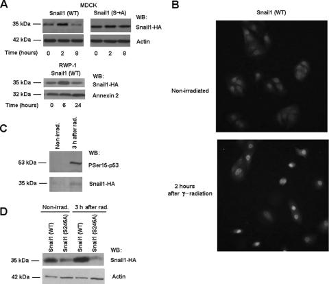
FIG. 7.
Snail1 protein is stabilized in response to γ radiation. Protein levels (A) and cellular distribution (B) of Snail1 were determined at the indicated times after irradiation in MDCK-Snail1 (wild-type [WT]), MDCK-Snail1 (Ser→Ala mutant [S→A]) or RWP-1-Snail1 cells by Western blotting (A) or immunofluorescence (B). Similar results were observed for another clone of MDCK-Snail1 cells. (C) Phosphorylation of Snail1 proteins was determined for stable MDCK-Snail1 transfectants 3 h after γ radiation (rad) and compared with the phosphorylation of nonirradiated (nonirrad) cells. Purification of phosphorylated Snail-HA was performed as indicated in Materials and Methods. (D) Snail1-HA protein levels in MDCK cells transiently transfected with wild-type or S246A Snail1-HA were determined as described above. The figure shows the result of a representative experiment of three (panels A and B for MDCK) or two (panel A for RWP-1 or panels C and D) performed. WB, Western blot.

