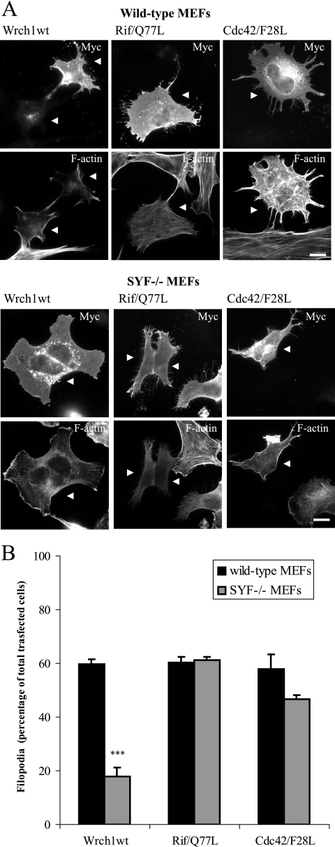
FIG. 8.
Src is required for the Wrch1-induced filopodia but not for the Rif- or Cdc42-induced filopodia. (A) Myc-tagged wild-type Wrch1 (Wrch1wt), Rif(Q77L), or Cdc42(F28L) was transfected into normal MEFs or MEFs isolated from SYF−/− mice. Myc-tagged Wrch1, Rif(Q77L), or Cdc42(F28L) was detected by a rabbit anti-Myc antibody followed by an AMCA-conjugated anti-rabbit antibody. Vinculin was detected by a mouse antivinculin antibody followed by a TRITC-conjugated anti-mouse antibody. Filamentous actin was detected by Alexa Fluor 488-conjugated phalloidin. The arrowheads mark transfected cells. Bar, 20 μm. (B) Quantification of the phenotypes observed in panel A was performed by microscopy analysis and scored as filopodium formation (filopodia) or stress fiber dissolution (SF loss). The values represent analyses of at least 100 transfectants in three independent experiments. The statistics analysis was carried out using Student's t test to compare the amounts of Wrch1-induced filopodia in wild-type and SYF−/− MEFs. * represents P values of <0.05, ** represents P values of <0.01, and *** represents P values of <0.001. The slightly reduced ability of Cdc42(F28L) to induce filopodia in SYF−/− MEFs is not statistically significant.

