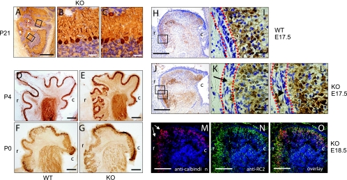
FIG. 2.
Development of malformations of the cerebellar cortex of nestin-Cre; Gna12−/−; Gna13flox/flox mice. (A to C) Sagittal section through the rostral part of a mutant cerebellum at P21 stained with cresyl violet and an anticalbindin antibody. The high-power images (B and C; boxes in panel A) show that despite the severe disorganization of the rostral cerebellar cortex, the basal lamination into an internal granule cell layer, Purkinje cell layer, and molecular layer was largely intact. (D to G) Sagittal sections of wild-type (D and F) and nestin-Cre; Gna12−/−; Gna13flox/flox (E and G) mice at P4 (D and E) and at P0 (F and G) stained with anticalbindin antibody. Sections are shown with the rostral part of the cerebellum facing the left side. (H to L) Sagittal sections of a wild-type (WT) (H and I) and a nestin-Cre; Gna12−/−; Gna13flox/flox (KO) cerebellum (J to L) at E17.5 stained with cresyl violet and an anticalbindin antibody. While the granule cell layer and the Purkinje cell layer are clearly separated in the wild type, clusters of Purkinje cells invade the external granule cell layer in mutant cerebella (arrow) and nests of Purkinje cells can be seen in the EGL (arrowhead). The EGL is marked by dotted red lines. (M to O) Sagittal section of a nestin-Cre; Gna12−/−; Gna13flox/flox cerebellum at E18.5 stained with anti-calbindin (red) and anti-RC2 antibodies (radial glia; green). Arrows in panel M indicate overmigrating Purkinje cells entering the EGL. r, rostral; c, caudal. Scale bars are 500 μm (A), 31.25 μm (B and C), 250 μm (D, E, H, J, and M to O), 125 μm (F and G), and 25 μm (I, K, and L).

