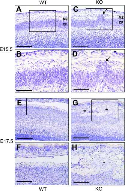
FIG. 3.
Development of cortical ectopia in nestin-Cre; Gna12−/−; Gna13flox/flox embryos. Shown are coronal sections of wild-type (WT) (A, B, E, and F) and mutant (KO) (C, D, G, and H) cortices at E15.5 (A to D) and E17.5 (E to H). Overmigration of cortical plate neurons in mutant cortices was first seen at E15.5 (arrow in C and D). At E17.5, huge areas of ectopic neurons had formed which filled the whole molecular layer and reached into the subarachnoidal space (indicated by stars in G and H). MZ, marginal zone; CP, cortical plate. Scale bars are 125 μm (A, C, F, and H), 62.5 μm (B and D), and 250 μm (E and G).

