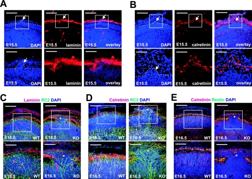
FIG. 4.
Immunohistochemical analysis of the cerebral ectopia in nestin-Cre; Gna12−/−; Gna13flox/flox embryos. (A and B) Cerebral cortices of E15.5 mice. nestin-Cre; Gna12−/−; Gna13flox/flox embryos were sectioned coronally and stained with antilaminin (A) or anticalretinin (B) antibodies. Sections were counterstained with DAPI. Shown are areas similar to those shown in Fig. 3C and D in which cortical plate neurons have invaded the molecular layer (arrows). Boxes indicate magnified areas, broken lines mark the outer border of the cortical plate. (C to E) Cerebral cortices of E16.5 wild-type (WT) and nestin-Cre; Gna12−/−; Gna13flox/flox (KO) embryos were sectioned coronally and stained with antilaminin (C, red), anti-RC2 (C and D, green), or anticalretinin (D and E, red) antibodies. Sections were counterstained with DAPI. Shown are representative areas of mutant cortices with ectopic neurons (marked by stars) and corresponding regions of cortices from wild-type embryos. Boxes indicate magnified areas. Bar lengths are 125 μm (upper panels in A to E), 41.5 μm (lower panels in A and B), and 62.5 μm (lower panels in C to E).

