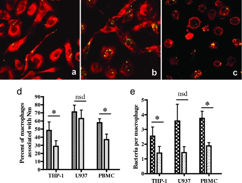
FIG. 6.
Uptake of CRP-opsonized and nonopsonized N. meningitidis by macrophages. Representative microscope images of PBMC-derived macrophages incubated with unopsonized GFP-expressing meningococci (a) and CRP-opsonized GFP-expressing meningococci (b) are shown. Macrophages were stained red with propidium iodide postinfection (a and b), or macrophage membranes were stained with a fluorescent lipophilic dye postinfection to enable z stacking through the macrophage to reveal internalized bacteria (c). (d and e) Enumeration of the association of bacteria with macrophages was carried out on at least 100 cells from 10 randomly selected fields of view for CRP-opsonized bacteria (cross-hatched bars) and unopsonized bacteria (stippled bars) colocalized with THP-1, U937, and peripheral blood-derived macrophages. The percentage of macrophages associated with bacteria was calculated (d) as well as the mean number of adherent or intracellular bacteria per macrophage (e). Data are means from at least three replicate experiments plus standard deviations (*, P < 0.05; nsd, no statistical difference).

