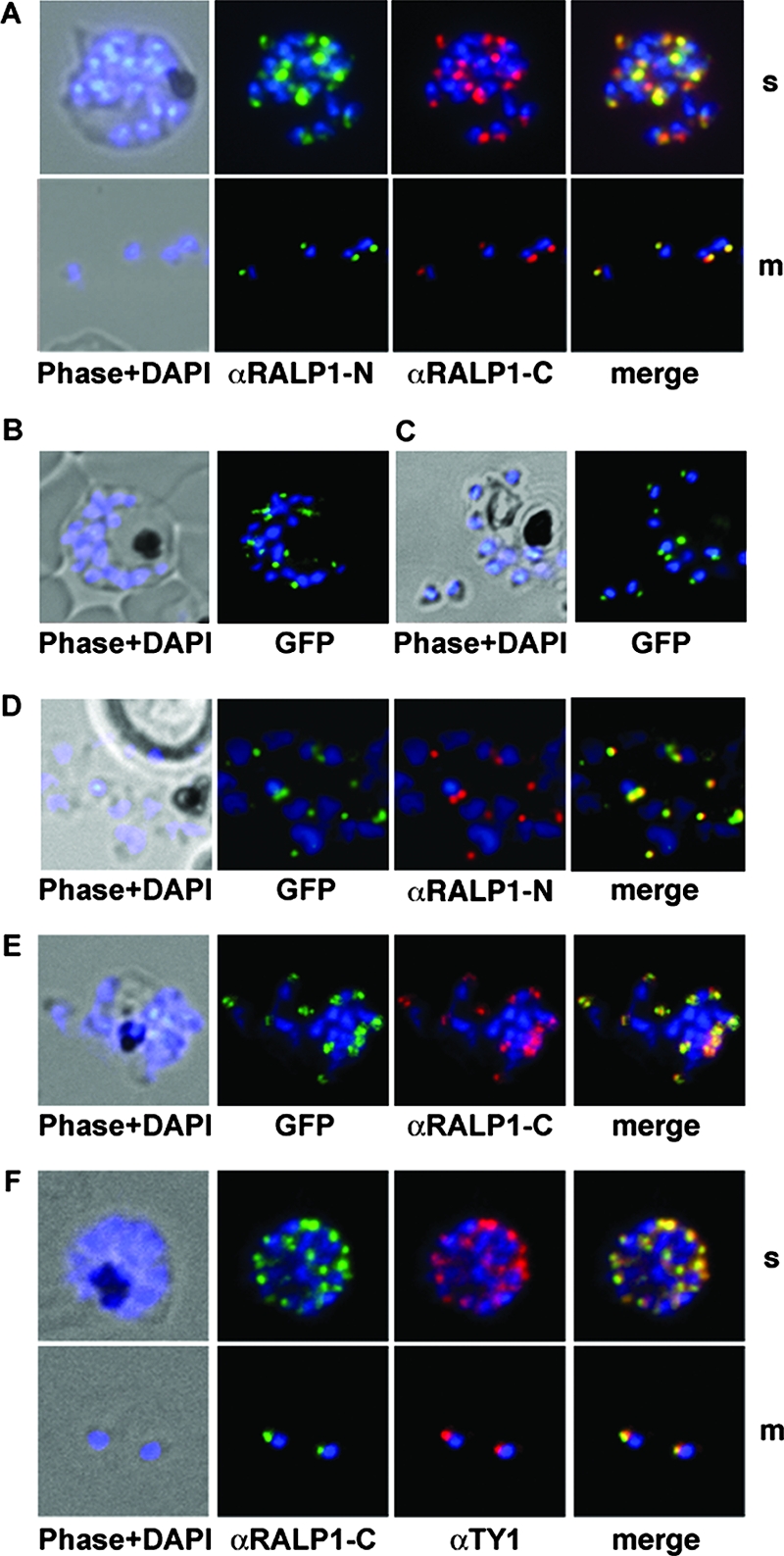
FIG. 4.

Subcellular distribution of RALP1-GFP fusion protein and immunofluorescence localization of RALP1 and RALP1-TY1. (A) Using methanol-fixed parasites, both RALP1-specific antibodies (αRALP1-N [green] and αRALP1-C [red]) localize the protein in the same subcellular compartment (merged image) in late schizonts (s) and free merozoites (m). The nucleus was stained with DAPI (blue). (B and C) Localization of RALP1-GFP by fluorescence microscopy in unfixed parasites in schizonts (B) and free merozoites (C). Using fluorescence of the GFP reporter protein in live cells, RALP1-GFP distribution (green) was restricted to the apical end of the parasite. The nucleus was stained blue. (D and E) Colocalization of RALP1-GFP with the endogenous RALP1 in free merozoites. N-terminal- and C-terminal-specific anti-RALP1 antibodies (αRALP1-N and αRALP1-C) (red) and the RALP1-GFP fusion protein (green) colocalize, as shown by the merged picture. (F) Colocalization of RALP1-TY1 with the endogenous RALP1 using RALP1-specific antibodies (αRALP1-C) (green) and anti-TY1 antisera (red) resulted in identical staining patterns at the apical end of the parasite.
