
FIG. 4.
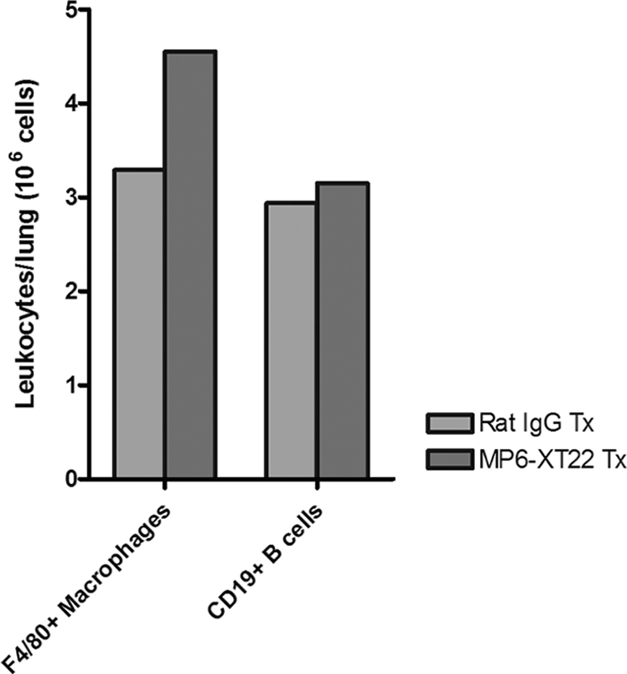
Quantification of F4/80+ macrophages and CD19+ B cells among CD45+ leukocytes obtained from lungs of MP6-XT22- and control rat IgG-treated mice with chronic tuberculosis through flow cytometry. Dead cells were identified and excluded from analysis by means of staining with the Live/Dead reduced biohazard cell viability kit number 2 (green staining) or number 4 (blue staining). Positively enriched live CD45+ cells were obtained and subjected to staining using antibodies for cell surface markers of interest. Absolute numbers of CD19+ B cells and F4/80+ macrophages at 9 days (9d) posttreatment are shown. Proportions of CD45+ cells analyzed were comparable between the two groups. Bars represent data from lung cells pooled from four or five mice. The experiments were repeated once with similar results.