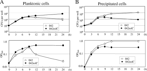
FIG. 6.
Time courses of the planktonic and precipitated cells in a microtiter plate assay. Strains were grown in triplicate in high-glucose DMEM in a 24-well microtiter plate at 37°C for 3 h, 6 h, 9 h, 12 h, and 24 h. (A) The planktonic cells in the medium were measured by plating serial dilutions on L-agar plates in triplicate (top). The concentration of planktonic cells in the medium was measured in terms of optical density at 600 nm (OD600) (bottom). (B) Spontaneously precipitated cells on the substratum after medium aspiration were collected by scraping into 500 μl of PBS, and viable cells were counted as described above (top) and the concentration was measured as described above (bottom). Data are presented as means of triplicate experiments, with error bars representing one SD. Where no error bars are visible, the deviation is smaller than the symbol.

