FIG. 4.
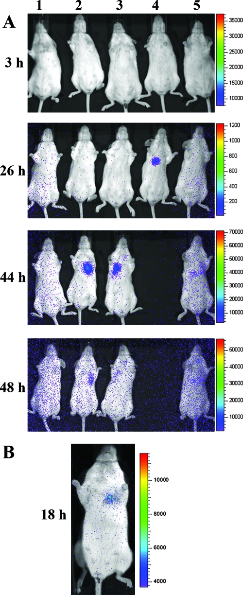
Merged luminescent and photographic images of mice after intranasal inoculation with ∼1 × 10⁷ B. anthracis spores transformed with pPS12. (A) Evidence of germination in the ventral thorax of three of five mice (animals 2, 3, and 4) within 48 h of infection. Mouse 5 appeared to have increased luminescence over the same area at 44 h, but the signal was weaker than with mice 2, 3, and 4. Mouse 4 died prior to the 48-h postinfection measurement. Note that for mice 2 and 3 the signal was on at 44 h and then off or decreased at 48 h, a pattern that is expected for a system that monitors spore germination but not vegetative cell outgrowth. Visualization of the low level of luminescence at 48 h was enhanced by increasing the background sensitivity, an adjustment that resulted in a generalized increase in background signal (blue dots) that is artificially concentrated around the periphery. (B) In a separate experiment, bioluminescence was detectable in one mouse at 18 h postinfection.

