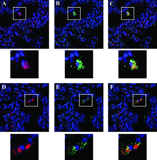
FIG. 7.
Immunohistochemistry of frozen sections of luminescent mouse lung tissue harvested 30 min after intranasal inoculation with spores of B. anthracis transformed with pPS12. (A to C) Tissue section 1. DAPI nuclear staining (blue) and anti-CD68 macrophage staining (red) show the presence of macrophages in the alveolar space and macrophages within the interstitial tissues (A); DAPI nuclear staining (blue) and anti-BclA spore staining (green) reveal spores in the alveolar space and in the tissues (B); merged images from panels A and B demonstrate spores in association with alveolar macrophages (C). (D to F) Tissue section 2. DAPI nuclear staining (blue) and anti-CD68 macrophage staining (red) again depict the presence of macrophages (D); DAPI nuclear staining (blue) and anti-EA-1 staining (green) illustrate that some vegetative antigen is in the lung at this early time point (E); the merged images from panels D and E show vegetative cell antigen colocalized with macrophages (F). Magnification (A to F), ×63. Inserts below each panel represent enlargement of the boxed area or cell in that panel.

