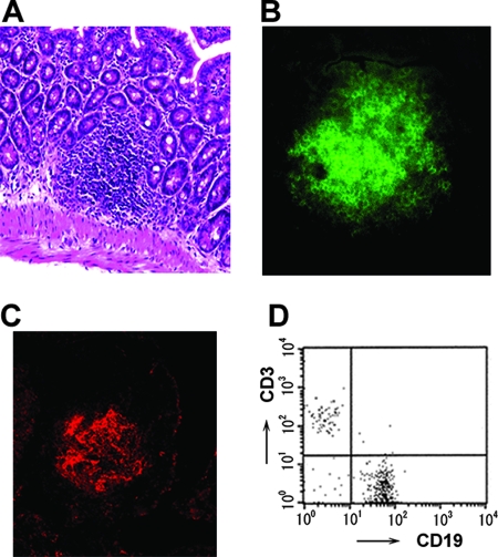
FIG. 1.
Characteristics of ILF in PP-null mice. (A) Hematoxylin-eosin staining. Immunohistochemical analyses of B cells (B) and FDC clusters (C) were performed by staining with FITC-anti-B220 Ab and PE-anti-CR1 Ab, respectively. Original magnification, ×100. Flow cytometric analysis shows the proportions of CD3+ T cells and CD19+ B cells (D).

