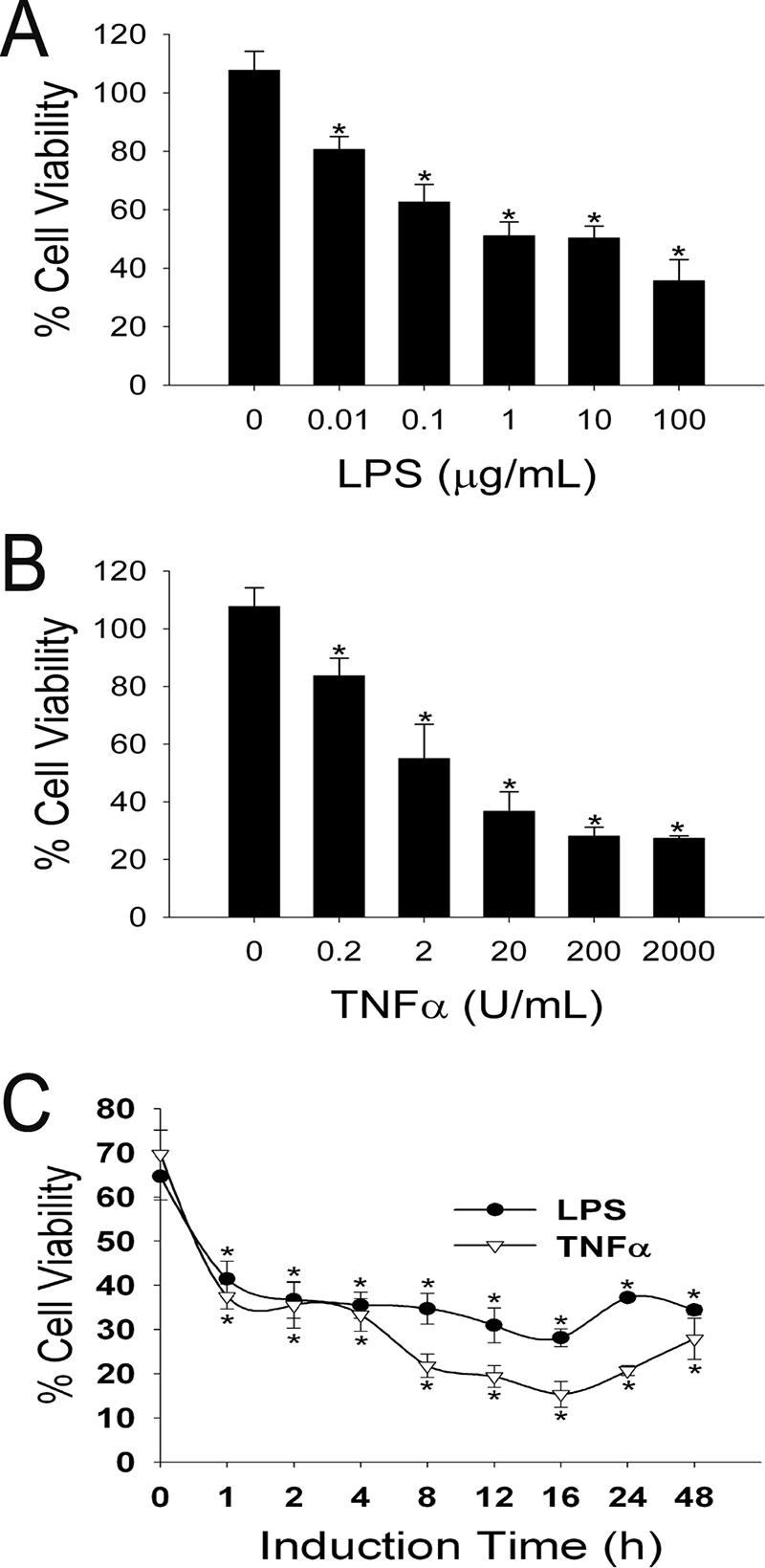
FIG. 1.

TNF-α and LPS induce Stx2 cytotoxicity in a dose- and time-dependent manner. (A and B) Confluent HUVEC were incubated with the indicated concentrations of (A) LPS or (B) TNF-α for 24 h before treatment with 1 nM Stx2 for 24 h. (C) Confluent HUVEC were incubated with either 10 μg/ml LPS or 200 U/ml TNF-α for the indicated times before treatment with 1 nM Stx2 for 24 h. Cell viability assays were performed, and the results were expressed as the percent viability for identical treatments for each concentration and time point without Stx2; the error bars indicate standard deviations (n = 4). An asterisk indicates that the value is statistically significantly (P < 0.01) different from the value for samples without LPS or TNF-α.
