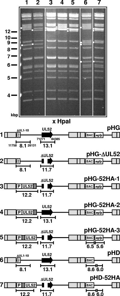
FIG. 1.
HCMV BAC genomes constructed in this study. The gel shows the HpaI restriction patterns of the recombinant BAC genomes. Relevant fragments are marked with white dots, and size markers are indicated to the left. The numbers of the lanes correspond to the genome structures shown below. (Bottom) Schematic drawing of the structures of the respective genomes. The sizes of fragments characteristic of each mutant are indicated. The nucleotide positions of HpaI restriction sites located around the UL1-10 and the UL52 locus are given in the first line (pHG). Gray boxes indicate repeat regions flanking the unique long and unique short genomic regions. The illustration is not drawn to scale.

