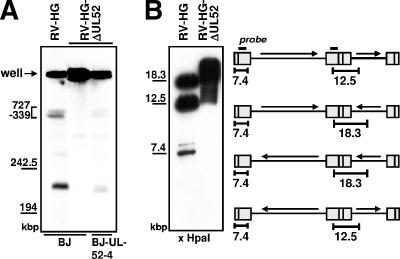
FIG. 4.
Viral concatemeric DNA is not cleaved in the absence of pUL52. (A) Normal BJ-1 cells were infected with the parental virus RV-HG or the UL52 deletion virus RV-HG-ΔUL52, and complementing BJ-UL52-4 cells were infected with RV-HG-ΔUL52 at an MOI of 1. Cells were harvested on day 5 p.i., embedded in agarose, and treated with proteinase K. Then, the DNA was resolved by pulsed-field gel electrophoresis and analyzed by Southern blotting using a probe specific for the a-repeat region. The positions of size markers derived from λ DNA are indicated to the left. (B) Normal BJ-1 cells were infected with the parental virus RV-HG or with the RV-HG-ΔUL52 mutant at an MOI of 1.5 and total DNA was isolated 5 days p.i. The DNA was cut with HpaI, followed by gel electrophoresis and Southern blotting using a probe derived from the b-sequence. The schematic drawing depicts the localization of the hybridization probe (black bar) as well as the DNA fragments corresponding to the internal repeat (12.5 and 18.3 kbp) and the left genomic terminus (7.4 kbp), depending on the four isoforms of the HCMV genome. The illustration is not drawn to scale.

